DOPS: bronchial blocker in a difficult airway, and a blocker that slipsDOPS: 58-year-old woman, previous neck radiotherapy, limited mouth opening
DOPS: bronchial blocker placement in a difficult airway, and dislodgement
From memory: the diagnosis and the notes are hidden. Answer each question aloud, then press Show.
Know this first
- Blockers suit difficult airways: keep the single-lumen tube, no tube exchange.
- Types: Arndt, Cohen, Fuji Uniblocker, EZ-Blocker; usually passed coaxially through the tube.
- Blockers dislodge more often than double-lumen tubes.
- Keep peak airway pressure below 30 cmH2O to stop gas leaking past the cuff.
- A cuff that slips into the trachea blocks ventilation and loses lung separation.
The patient
The examiner asks
Answer aloud, then press Show.
Prepare
How do you prepare?
Show the answerHide the answer
- Difficult airway, so I keep her single-lumen tube and add a blocker instead of exchanging tubes. (Procedure step)
- Options: Arndt, Cohen, Fuji Uniblocker or EZ-Blocker; I'll pass an Arndt through the tube.
- Multiport adapter, lubricated fibreoptic scope, blocker cuff tested, suction ready.
Perform
Talk me through placement.
Show the answerHide the answer
- Blocker through the adapter, with its wire loop around the fibreoptic scope. (Procedure step)
- Scope into the left main bronchus; slide the blocker along it, then retract the loop.
- Inflate the cuff under direct vision until it seals the left main bronchus.
- Lock the blocker at the adapter and secure the tube firmly.
Perform
How do you confirm isolation and keep it?
Show the answerHide the answer
- Scope check: the inflated cuff sits in the left main bronchus, clear of the carina. (Procedure step)
- Suction through the blocker lumen helps the left lung deflate.
- Recheck with the scope after turning her lateral, since blockers shift easily.
- Keep peak airway pressure under 30 cmH2O so gas doesn't leak past the cuff.
Crisis
During surgical handling, ventilation suddenly fails: peak pressure 45 cmH2O, capnography trace flattening, SpO2 92% and falling. What do you do?
Show the answerHide the answer
- The blocker cuff has probably slipped into the trachea and is obstructing both lungs. (Crisis)
- Deflate the blocker cuff at once and ventilate with 100% oxygen.
- Tell the surgeon to pause lung retraction.
- When saturation recovers, scope down the tube and reposition the blocker into the left bronchus.
- Reinflate under vision, lock it again, and recheck after any further movement.
Scan · Bronchoscopy: right upper lobe
Here is your image. Point to and name each structure, then say why it matters.
Fibreoptic bronchoscope via the tracheal lumen, then through the right DLT's upper-lobe slot.
Inside the right upper lobe bronchus (per caption); scope rotation not stated
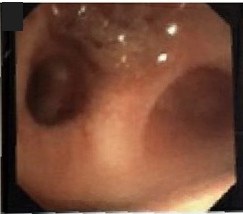 , open the image full size
Show the answerHide the answer
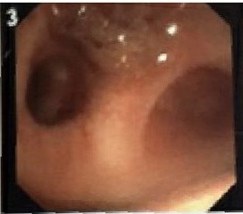 , open the image full size
This image carries no labels of its own; the structures are named below.
Labels Right upper lobe (from the source caption)
- On the right side wall, this opening is the right upper lobe.
- Going in shows three holes, which only this lobe has.
- It starts about two centimetres below the carina, sometimes higher, even off the trachea.
- A right tube's slot must line up here, or the lobe is cut off.
- Right-sided blockers have the same small margin for error.
- Right upper lobe orifice Lateral wall, about 3–4 o'clock, roughly 2 cm below carina
- Three segmental openings Apical, anterior, posterior: unique trifurcation that identifies it
- Anomalous take-off May be under 2.5 cm away or arise from trachea
- Right DLT ventilation slot Must sit over the orifice; margin only 1–8 mm
- Bronchus intermedius Seen beyond the slot through the bronchial lumen
Checked against: Miller 10e · Barash 9e
Image: Knight SE et al., Pigtail catheter found in the Lingula of a pediatric patient on VV-ECMO while performing flexible bronchoscopy: a case report on an unexpected finding (Front Med (Lausanne) 2026; doi:10.3389/fmed.2026.1912185), Figure 2 (image 3, cropped), CC BY 4.0. Changes: Trimmed the frame edge. Question image: panel number '3' covered
Scan · Bronchoscopy: bronchial blocker
Here is your image. Point to and name each structure, then say why it matters.
Fibreoptic bronchoscope through the single-lumen tube, viewing from just above the carina.
From above the carina: blocker (yellow shaft) with inflated balloon in the left main bronchus at upper left, open right main bronchus below
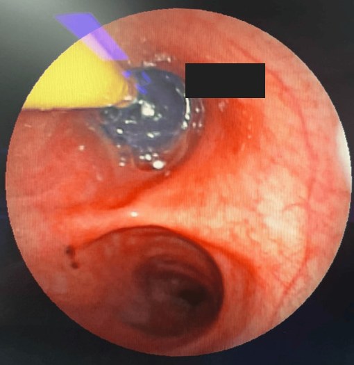 , open the image full size
Show the answerHide the answer
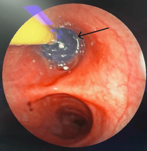 , open the image full size
Labels Arrow: the blocker with its balloon inflated
- From just above the carina, the blocker goes into the chosen main bronchus.
- I inflate the balloon while watching, using only enough to seal.
- The balloon stays below the carina, not bulging into the trachea.
- On the right there is less room because the upper lobe comes off early.
- If it slips into the trachea we cannot ventilate, so I deflate it immediately.
- Main carina Viewed from just above it in the trachea
- Blocker balloon in main bronchus Inflate under vision with least volume that seals
- Balloon edge clear of carina Must not ride up over the carina
- Right-sided placement Upper lobe close by: smaller margin for error
- Dislodged balloon in trachea Blocks both lungs: hypoxia, arrest; deflate at once
Checked against: Miller 10e · Barash 9e
Image: Gupta S et al., Anesthetic Management for the Retrieval of Retained Intrathoracic Foreign Bodies Adjacent to Critical Mediastinal Structures Using One-Lung Ventilation With a Bronchial Blocker: A Report of Two Cases (Cureus 2026; doi:10.7759/cureus.110538), Figure 7, CC BY 4.0. Changes: Cropped to the round scope image. Question image: black arrow covered with a box on the bronchial wall
Fatal mistakes
- Placing the blocker blind.
- Leaving the cuff inflated when ventilation fails.
Checked against: Miller 10e · Barash 9e
Report a problem with this case
Mark Got it only if you said every point. Again brings the case back tomorrow; Got it brings it back in 3, then 7, 14 and 30 days.