DOPS: ultrasound-guided internal jugular line, wire ectopy and air embolismDOPS: 66-year-old man, ventilated in ICU with septic shock, on noradrenaline via a peripheral cannula
DOPS: ultrasound-guided internal jugular line, wire ectopy and air embolism
From memory: the diagnosis and the notes are hidden. Answer each question aloud, then press Show.
Know this first
- Ultrasound guidance is now standard for internal jugular access.
- Maximal barrier precautions and chlorhexidine–alcohol prep reduce line infection.
- Confirm venous placement by manometry or pressure before dilating; verify the tip position.
- Arterial puncture is commoner than pneumothorax.
- Air embolism: stop entry, 100% oxygen, left side down and head down, aspirate the line.
The patient
The examiner asks
Answer aloud, then press Show.
Prepare
How do you prepare?
Show the answerHide the answer
- He lacks capacity: consent from his guardian, or the emergency two-doctor route if none is reachable. (Procedure step)
- Clotting is near normal: INR 1.3, platelets 140.
- Maximal barrier: hat, mask, sterile gown and gloves, and a full-length drape.
- Chlorhexidine–alcohol skin prep; it lowers line infection compared with povidone–iodine.
- Pre-scan: compressible vein, pulsatile carotid, and a patent vein without clot.
Perform
Talk me through insertion.
Show the answerHide the answer
- Real-time ultrasound, keeping the needle tip in view until it is inside the vein. (Procedure step)
- Wire through the needle, then scan to see the wire inside the vein, not the artery.
- Before dilating, confirm venous placement by manometry or a pressure trace.
- Dilate, railroad the catheter, never letting go of the wire, then remove it intact.
- Aspirate, flush and cap every lumen; confirm the tip position before use.
Crisis
As the wire is advanced, runs of ventricular tachycardia appear. BP 84/50. What do you do?
Show the answerHide the answer
- The wire is irritating the heart: pull it back until the rhythm settles. (Crisis)
- Check for a pulse; pulseless VT means starting the arrest algorithm.
- If VT persists with a pulse and low BP: synchronised cardioversion.
- Re-advance only to the depth needed, watching the monitor.
Crisis
Later a hub is found open with the bed head-up. BP 70/40, HR 125, EtCO2 falls from 4.8 to 2.5 kPa, SpO2 88%.
Show the answerHide the answer
- Venous air embolism: close the open hub immediately. (Crisis)
- 100% oxygen.
- Tilt him head-down, left side lowest, to move air out of the right ventricular outflow.
- Aspirate air back through the central line.
- Support the circulation with fluid and vasopressors; CPR if he arrests.
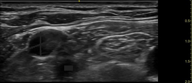
Scan · Internal jugular vein and carotid
Here is your image. Point to and name each structure, then say why it matters.
Real-time imaging across the vessel (short axis), or along it as an option (probe type and position not given in the corpus).
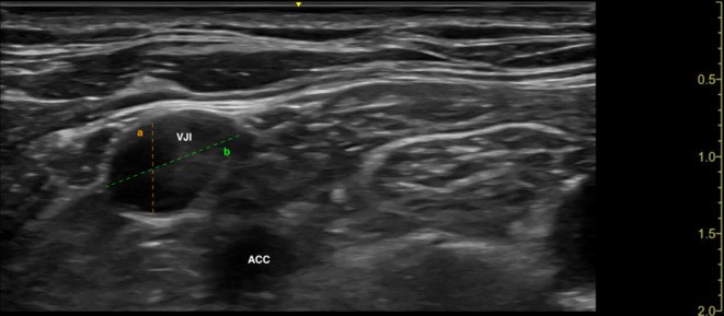
Transverse short axis, skin at top; IJV superficial and to the left, common carotid deeper and to the right; side of neck not stated
 , open the image full size
Show the answerHide the answer
 , open the image full size
Labels VJI internal jugular vein · ACC common carotid artery
- I scan before prepping to check the vein is a good size and not clotted.
- This is the internal jugular; the right side is my usual choice.
- This is the carotid, the thing I must not puncture.
- I keep the beam just ahead of the tip until it sits inside the vein.
- Before dilating I check pressure to exclude arterial placement.
- Right internal jugular vein Preferred side: predictable anatomy, reachable from the head end
- Carotid artery Accidental puncture is the classic danger; confirm venous by manometry
- Vein size and patency Pre-scan rules out a small or clotted vein
- Needle tip in lumen Follow the tip ahead of time until it sits mid-vessel
- Back wall of the vein Long-axis view helps avoid going through it
Checked against: Barash 9e · Miller 10e
Image: Kalina M et al., A novel "lateral approach short axis in-plane" technique vs. conventional "short-axis out-of-plane approach" for ultrasound-guided internal jugular vein access: a prospective randomized non-inferiority trial (Ultrasound J 2025; doi:10.1186/s13089-025-00405-9), Fig. 2, CC BY 4.0. Changes: Whole image kept, no crop. Question image: orange and green measurement lines and their 'a'/'b' letters covered with dark grey along the lines; 'VJI' and 'ACC' labels covered with boxes
Fatal mistakes
- Dilating the artery.
- Losing the guidewire.
- Leaving a hub open with the patient upright.
Checked against: Barash 9e · Miller 10e · Saudi Guidelines for Informed Consent, First Edition 1440H (2019)
Report a problem with this case
Mark Got it only if you said every point. Again brings the case back tomorrow; Got it brings it back in 3, then 7, 14 and 30 days.