DOPS: left double-lumen tube and desaturation on one lungDOPS: a 62-year-old man, 176 cm, 80 kg, for elective thoracoscopic right upper lobectomy
From memory: the diagnosis and the notes are hidden. Answer each question aloud, then press Show.
Know this first
- Size: men over 170 cm 41 Fr; under 170 cm 39 Fr; women 37 or 35 Fr.
- Left DLT depth at the teeth: about 12 + height/10 cm.
- Auscultation alone missed malposition in 48% on fibreoptic check.
- One-lung ventilation: tidal volume 4–6 mL/kg IBW, PEEP, recruitment; FiO2 1.0 if desaturating.
- Most desaturation comes early, in the first 10 minutes on one lung.
The patient
The examiner asks
Answer aloud, then press Show.
Prepare
How do you prepare?
Show the answerHide the answer
- Right lobectomy, so a left-sided tube; it avoids blocking the right upper lobe bronchus. (Procedure step)
- He's a man over 170 cm, so a 41 Fr tube.
- Depth at the teeth about 12 plus height over 10: roughly 29 to 30 cm.
- Test both cuffs, lubricate, and have the fibreoptic scope, connector and clamps ready.
Perform
Talk me through insertion and confirmation.
Show the answerHide the answer
- Bronchial tip through the cords, turn 90 degrees anticlockwise, advance gently without force. (Procedure step)
- Tracheal cuff up, ventilate, check both sides move and sound equal.
- Clamp each limb in turn: the clamped side stops moving, the other side keeps ventilating.
- Scope down the tracheal lumen: blue bronchial cuff just below the carina.
- Scope down the bronchial lumen: the left upper lobe opening is clear.
- After turning him lateral, I recheck with the scope, as tubes often move.
Perform
How do you set up one-lung ventilation?
Show the answerHide the answer
- Two lungs for as long as possible; isolate the right lung when the surgeon needs it. (Procedure step)
- Tidal volume 4–6 mL/kg of his ideal weight, with PEEP and recruitment breaths.
- Keep plateau pressure under 25 cmH2O and adjust the rate for carbon dioxide.
- Watch saturation closely; most falls come within the first 10 minutes.
Crisis
Five minutes into one-lung ventilation, SpO2 falls from 97% to 86%. BP 125/75, HR 80. What do you do?
Show the answerHide the answer
- Desaturation on one lung: FiO2 to 1.0 and tell the surgeon. (Crisis)
- Scope the tube: has it moved, or is a lobe of the left lung blocked?
- Check blood pressure and cardiac output; keep volatile below 1 MAC.
- Recruit the ventilated left lung, then increase its PEEP.
- Give the right lung CPAP of 1–2 cmH2O after a recruitment breath, if surgery allows.
- If it falls fast or keeps falling, return to two-lung ventilation.
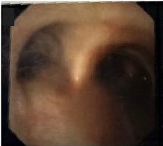
Scan · Bronchoscopy: the carina
Here is your image. Point to and name each structure, then say why it matters.
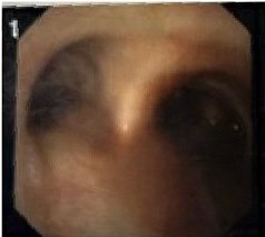
Looking down the trachea onto the main carina; scope rotation not stated
 , open the image full size
Show the answerHide the answer
 , open the image full size
This image carries no labels of its own; the structures are named below.
Labels The main carina (from the source caption)
- Cartilage rings are in front; the smooth flat wall is at the back.
- This ridge ahead is the main carina.
- The wider, straighter opening is the right main bronchus.
- Just inside it, on the side wall, this hole with three openings is the right upper lobe.
- The other side, longer before it divides, is the left main bronchus.
- Tracheal rings and posterior wall Rings in front, flat membranous wall behind: sets orientation
- Main carina Sharp ridge where the trachea splits
- Right main bronchus Wider, straighter, short before it branches
- Right upper lobe take-off On the right lateral wall, three openings: proves it is right
- Left main bronchus Longer, about 5 cm, before its first split
Checked against: Barash 9e · Miller 10e
Image: Knight SE et al., Pigtail catheter found in the Lingula of a pediatric patient on VV-ECMO while performing flexible bronchoscopy: a case report on an unexpected finding (Front Med (Lausanne) 2026; doi:10.3389/fmed.2026.1912185), Figure 2 (image 1, cropped), CC BY 4.0. Changes: Trimmed the frame edge. Question image: panel number '1' covered
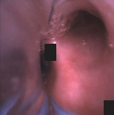
Scan · Bronchoscopy: left double-lumen tube
Here is your image. Point to and name each structure, then say why it matters.
Fibreoptic bronchoscope down the tracheal lumen first, then the bronchial lumen; repeat after repositioning.
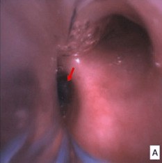
Down the tracheal lumen of a left DLT onto the carina; scope rotation not stated
 , open the image full size
Show the answerHide the answer
 , open the image full size
Labels Arrow: bronchial cuff of the left double-lumen tube in the left main bronchus
- Down the tracheal lumen I see the carina straight ahead.
- The blue cuff edge sits just inside the left bronchus, below the carina.
- It does not bulge over the carina or push it right.
- On the right I find the upper lobe with its three openings.
- Down the bronchial lumen, both left lobar openings are clear.
- I recheck with the scope after every position change.
- Tracheal carina Seen straight ahead down the tracheal lumen
- Blue bronchial cuff edge In left bronchus, about 5–10 mm below carina
- No cuff herniation Bulging cuff can block the right main bronchus
- Right upper lobe take-off Confirms which side you are looking at
- Left upper and lower lobe orifices Down bronchial lumen: both open, none blocked or impacted
Checked against: Miller 10e · Faust · Barash 9e
Image: Tan DJA et al., Difficult Lung Isolation in a Heavy Smoker With a History of Left Lower Lobectomy: A Case Report (Cureus 2024; doi:10.7759/cureus.69388), Figure 2, CC BY 4.0. Changes: Cropped to panel A. Question image: red arrow and panel letter A covered with boxes
Fatal mistakes
- No fibreoptic confirmation.
- Persisting with one-lung ventilation while hypoxia worsens.
Checked against: Miller 10e · Barash 9e
Report a problem with this case
Mark Got it only if you said every point. Again brings the case back tomorrow; Got it brings it back in 3, then 7, 14 and 30 days.