DOPS: ultrasound-guided popliteal sciatic blockDOPS: a 48-year-old woman, 70 kg, for open fixation of an ankle fracture
DOPS: ultrasound-guided popliteal sciatic block and intraneural injection
From memory: the diagnosis and the notes are hidden. Answer each question aloud, then press Show.
Know this first
- Sciatic splits into tibial and common peroneal branches near the popliteal fossa.
- Ideal site: at or just above the bifurcation, about 6–10 cm above the crease.
- In-plane needle; tip in the gap between tibial and peroneal branches.
- Injection pressure above 15 psi can damage nerves; never inject against resistance.
- Ropivacaine maximum 3 mg/kg.
The patient
The examiner asks
Answer aloud, then press Show.
Prepare
How do you prepare?
Show the answerHide the answer
- Stop before I block: confirm identity, consent, the side and the surgical site mark. (Procedure step)
- Monitoring, IV access, resuscitation drugs and lipid emulsion at hand.
- She stays awake or lightly sedated, so she can tell me if injection hurts.
- Ropivacaine, staying well below the 3 mg/kg maximum.
- Supine with the leg raised; probe behind the knee.
Perform
Talk me through the block.
Show the answerHide the answer
- Linear high-frequency probe at the crease: tibial and common peroneal nerves beside the vessels. (Procedure step)
- Scan upwards until they join as the sciatic nerve, about 6–10 cm above the crease.
- In-plane from the lateral side, needle tip always in view.
- Tip between the tibial and peroneal branches at the split, inside the shared sheath.
- Aspirate, then inject in small aliquots, watching spread around both nerves.
- About 20–30 mL in total, aspirating before each aliquot.
Perform
After the injection, what do you do?
Show the answerHide the answer
- Test sensation and movement of the foot before she goes to theatre. (Procedure step)
- Record the drug, volume, approach, image findings and any events.
- Warn her the foot will stay numb for hours, so she must protect it.
- Review her the next day and ask about lasting numbness or weakness.
Crisis
On injection the syringe becomes hard to push and she cries out with sharp pain shooting to her foot.
Show the answerHide the answer
- Possible intraneural injection: stop injecting immediately. Crisis
- Pressure above 15 psi can injure the nerve; I never push against resistance.
- Withdraw the needle slightly and recheck the image; the tip must be outside the nerve.
- Inject a small test volume; continue only if it flows easily and without pain.
- Document the event, tell her, and review her the next day for numbness or weakness. After surgery
Scan · Sciatic nerve at the popliteal fossa
Here is your image. Point to and name each structure, then say why it matters.
Linear 10–15 MHz probe transverse in the popliteal fossa, starting at the crease and scanning upward.
Transverse at the popliteal fossa, at the bifurcation
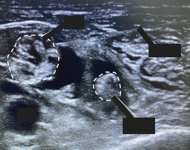 , open the image full size
Show the answerHide the answer
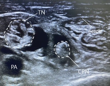 , open the image full size
Labels TN tibial nerve · CPN common peroneal nerve · PA popliteal artery · needle
- At the crease these pulsing circles are the popliteal vessels.
- Above and lateral to them are the tibial nerve and, furthest out, the common peroneal.
- Scanning up, they merge into this large bright oval, the sciatic nerve.
- This muscle on top is biceps femoris, and deep is the femur.
- I want a doughnut of drug around the nerve just before it divides.
- Popliteal artery and vein Doppler landmark at the crease; nerves lie above and lateral
- Tibial nerve Next to the vessels at the crease; drifts lateral going up
- Common peroneal nerve The most lateral nerve
- Sciatic nerve (junction) Big bright oval; ideal site before the split, 6–10 cm up
- Biceps femoris Lies over the nerves higher in the thigh
- Femur Bright bony border deep in the image
Checked against: Barash 9e
Image: Yoo SW et al., Comparing quality of recovery and satisfaction between spinal anesthesia and nerve block in orthopedic below-knee surgery: A prospective controlled trial (Medicine (Baltimore) 2024; doi:10.1097/md.0000000000037573), Figure 1 (panel A, cropped), CC BY 4.0. Changes: Cropped the figure letter; in the question image the TN, CPN, PA and needle labels and their leader lines are covered; the dashed nerve outlines are kept.
Fatal mistakes
- Injecting against high pressure.
- Blocking a deeply sedated patient.
Checked against: Miller 10e · Barash 9e · Implementation Manual: WHO Surgical Safety Checklist (1st edition, Safe Surgery Saves Lives) (2009)
Report a problem with this case
Mark Got it only if you said every point. Again brings the case back tomorrow; Got it brings it back in 3, then 7, 14 and 30 days.